Table of Contents
Link to: 2.2.5.2.5 Methanol Teaching Resources
Link to: Methanol Problems
Methanol
OVERVIEW
Methanol (methyl alcohol, wood spirits) is used as a fuel in high performance engines, solvents, as an antifreeze or window cleaner. Methanol is also used to fortify illicit spirits. Methanol is a colourless, flammable solution with a slightly alcoholic smell. Almost all significant exposures occur via ingestion, but methanol can also be absorbed via inhalation and through the skin. Small amounts of methanol are ingested with foods such as fresh fruit juices and vegetables. Methanol is a natural fermentation product found in all spirits. Serum methanol concentrations rise following binge drinking and are thought to be one cause of a hangover.
Methanol toxicity produces a raised anion-gap metabolic acidosis, ocular and central nervous system toxicity. Treatment with an alcohol dehydrogenase inhibitor such as ethanol or fomepizole may be life-saving.
TOXICITY
Fifiteen mL of a 40% solution has been fatal, and as little as 10mL has caused blindness. Due to the current relative concentrations of methanol (2%) and ethanol (98%) in methylated spirits it is the ethanol which is the more toxic in acute ingestions. In Australia there is no methanol contained in methylated spirits.
MECHANISM OF TOXIC EFFECTS
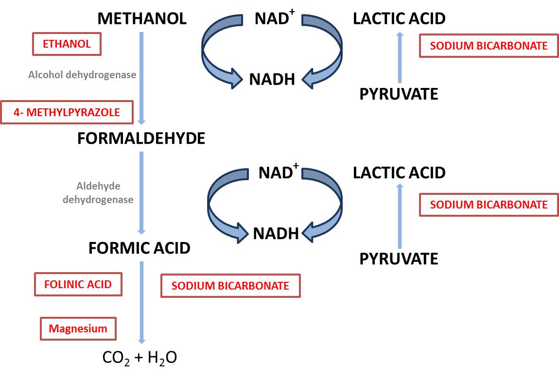
Methanol is metabolised by alcohol dehydrogenase to formaldehyde, which does not accumulate to a significant degree, but is metabolised to formic acid. Formic acid is metabolised more slowly and accumulates. Formic acid dissociates to formate and a hydrogen ion. The formate is metabolised to CO2 and water by a folate-dependent mechanism (Figure 1).
Formic acid inhibits mitochondrial cytochrome oxidase activity, preventing oxidative metabolism and so producing “tissue hypoxia” in a manner similar to cyanide and carbon monoxide.
Systemic acidosis is produced through the production of formic acid and the generation of a lactic acidosis because of inhibition of cellular aerobic metabolism. Worsening acidosis promotes the generation of further undissociated formic acid and the movement of this entity across cellular membranes into the central nervous system. Further CNS depression and hypotension lead to further lactic acid production and a downward spiral of worsening acidosis.
Formation of formic acid is the most likely cause of the early raised anion-gap metabolic acidosis seen following methanol exposure. Consequent interruption of normal cellular respiration by formic acid then leads to the production of a lactic acidosis (Figure 2).
Figure 1. Metabolism of methanol
Figure 2. Mechanism of acidosis following methanol exposure
Ocular toxicity is caused by undissociated formic acid, which directly targets the optic disc and retrolaminar section of the optic nerve. Acidosis worsens the ocular toxicity by promoting movement of formic acid into cells and decreasing the amount of undissociated formic acid. Optic disc oedema, breakdown of myelin sheaths and optic nerve lesions result.
Central nervous system findings demonstrated on CT, MRI and at autopsy following significant methanol poisoning include oedema and necrosis of the basal ganglia (particularly the putamen) and haemorrhages in the subcortical white matter. The putamen is known to have high rates of oxygen and glucose consumption and for this reason is thought to be more vulnerable to the adverse effects of formic acid.
KINETICS IN OVERDOSE
Absorption
Methanol is rapidly absorbed from the gastrointestinal tract with peak concentrations occurring within 30-60 minutes. Methanol is relatively well absorbed via the skin and respiratory system.
Distribution
Methanol is water soluble and has a volume of distribution similar to body water (0.6 L/kg).
Metabolism - Elimination
The major pathway is metabolism by alcohol dehydrogenase to formaldehyde. Formaldehyde is then oxidised to formic acid by formaldehyde dehydrogenase. In the poisoned patient methanol elimination is best characterised by zero order kinetics. The apparent elimination half-life is approximately 24 hours. At low concentrations first-order kinetics are apparent with a half-life of 1-3 hours.
CLINICAL EFFECTS
Methanol (as compared with ethanol) causes relatively little initial inebriation. Following ingestion there is an asymptomatic period (6-36 hours, up to 90 hours if ethanol co-ingested) while methanol is being metabolised into its toxic metabolite formic acid. This period is generally shorter with increasing dose and will be longer in those individuals who have co-ingested ethanol. Visual disturbance with a clear sensorium is suspicious for methanol poisoning. Following significant exposure a range of symptoms and clinical signs ensue:
Visual symptoms commonly include blurred vision, decreased acuity and the feeling of being in a snow field. Clinical findings include hyperaemia of the optic disc and reduced pupillary responses to light. Permanent optic nerve damage occurs in 25% of survivors. Parkinsonism and necrosis of the putamen have also been reported.
INVESTIGATIONS
Anion gap
Metabolism of methanol and the formation of formic and lactic acids produces a raised anion gap metabolic acidosis. It is important to remember that patients who present early following methanol ingestion, prior to significant methanol metabolism, may have normal acid-base status and a normal anion gap. This situation signifies a good prognosis, as long as the diagnosis is made and appropriate treatment instituted to prevent the formation of toxic metabolites. Patients who present late following significant methanol ingestion are likely to have an established raised anion-gap metabolic acidosis.
Osmolal gap
There are many pitfalls in using the osmolal gap to make the diagnosis of significant methanol exposure. An ethanol concentration is required to calculate a meaningful osmolal gap in methanol poisoning. Ethanol will contribute to any osmolal gap. A significantly raised osmolal gap is useful in providing evidence to confirm a significant methanol exposure, but a normal osmolal gap is not reassuring and does not exclude the diagnosis for a number of reasons:
1. Many patients will have a negative resting osmolal gap. A toxic exposure of methanol may not raise the osmolal gap above the commonly quoted normal upper limit of +10 mOsmol/kg H20.
2. Patients who present late after significant methanol exposure following metabolism of the parent molecule will have a minimal increase in the osmolal gap (as methanol is the primary contributor to the increased measured osmolality). In this case the osmolal gap is likely to be normal, but there will be a significantly raised anion-gap metabolic acidosis.
Biochemistry
Electrolytes, renal function and glucose should be monitored. A serum ethanol concentration is required to interpret the osmolal gap and serial serum ethanol concentrations obtained in a near real-time manner are required to effectively use ethanol as an antidote. Acid-base status should be monitored using venous blood gas analyses. Patients who are intubated or have respiratory compromise will require arterial blood gas monitoring. Patients with a history of alcohol abuse may be hypomagnesaemic or hypokalaemic.
Serum methanol concentrations
Conversion factor:
* g/L x 31.3 = mmol/L
- mmol/L x 0.032 = g/L
A quantitative serum concentration of methanol may be useful to indicate the need for haemodialysis. Availability of serum methanol concentrations is often an issue in suspected cases of ingestion, and often the diagnosis has to be made on the basis of history and collaborative biochemical evidence. A peak methanol concentration of > 50 mg/dL is associated with severe toxicity. A peak of less than 20 mg/dL is not associated with severe toxicity, but this concentration is only reassuring in a clinically well patient without a metabolic acidosis (a low methanol concentration may be measured in late presenters following metabolism of the parent molecule).
See the following for lethal concentrations of alcohols and their corresponding osmolal gaps:
Substance: Ethanol
Molecular Weight: 46
Potentially Lethal Concentration (mg/dL): 350
Corresponding Osmolal Gap (Osmol/kg H2O): 75
Substance: Methanol
Molecular Weight: 32
Potentially Lethal Concentration (mg/dL): 80
Corresponding Osmolal Gap (Osmol/kg H2O): 25
Contribution of Alcohol to Osmolar Gap
Osmolal gap=(Ethanol [mg/dL])/3.7 – 0.35
or, in SI units:
Osmolal gap (Osmol/kg)=1.25 (Ethanol [mmol/L]) – 0.35
(from Purssell et al)
DIFFERENTIAL DIAGNOSIS
Methanol is one of a number of drugs that can lead to an unconscious patient with a metabolic acidosis. Visual disturbance and a raised anion gap make the diagnosis more likely. A normal osmolal gap does not exclude significant methanol poisoning but a raised osmolal gap is very suggestive of poisoning with methanol, ethylene glycol, or other toxic alcohols and ethers.
DETERMINATION OF SEVERITY
Ingested dose
A dose in an adult of as little as 60 mL may cause significant toxicity or death. Ocular toxicity has been reported following ingestions of 20-30 mL.
Prognostic features
The following clinical signs are associated with a poor prognosis and are an indication for intensive care admission and usually haemodialysis.
- Coma
- Visual disturbance
- Hypotension
- Methanol concentration > 50 mg/dL
- Severe acidosis (pH < 7.1)
MANAGEMENT
Initial management should be directed at stabilising airway, breathing and circulation. Treatment is then directed at limiting or preventing acidosis, ophthalmological damage and coma.
Supportive
Patients should have IV access with generous IV. Patients with established clinically significant poisoning should be managed in a critical care setting. Patients with a significant methanol ingestion who present early without an established metabolic acidosis may potentially be managed in a ward environment if fomepizole is used as an antidote. Outcome is correlated with the degree of acidosis at the time of presentation. A pH below 7.3 should be corrected with intravenous sodium bicarbonate to obtain a pH in the normal range (7.35-7.45). Seizures should be treated with benzodiazepines.
GI Decontamination
Methanol is very rapidly absorbed and is not well adsorbed to charcoal. In most patients therefore GIT decontamination will not be helpful.
Antidotes
Traditionally ethanol has been the mainstay of treatment. However if available fomepizole should be first line treatment.
Ethanol
Ethanol has a higher affinity for alcohol dehydrogenase than methanol (20-30x) and competitively inhibits the metabolism of it to more toxic metabolites. Thus, it is most useful prior to the conversion of methanol to toxic metabolites and has little role in patients with low methanol concentrations and a marked acidosis. Ethanol is indicated for significant methanol ingestion or a methanol concentration > 20 mg/dL. Ethanol should be given orally or intravenously if the oral administration is not tolerated. A blood ethanol of 100 mg/dL (0.1 g/dL (%), 21.7 mmol/L) is required to maximally inhibit alcohol dehydrogenase.
Loading dose
Loading dose = Cp (target concentration) * Vd (volume of distribution)
= 1 g/L * 0.6 * body weight (kg)
= 0.6 g * body weight
Thus for a 60 to 70 kg adult the loading dose is 36 - 42 g of ethanol. This is equivalent to:
- 4 standard drinks (4 * 30 mL of spirits)
- or
- 360 to 420 mL of 10% ethanol IV
Maintenance dose
The dose required to maintain this concentration is 5 - 10 g/hour (depending on whether the enzymes in the patient have been induced by chronic alcohol consumption). This rate should be doubled during dialysis. Ethanol concentrations should be monitored and the rate adjusted accordingly.
Fomepizole
This is an alcohol dehydrogenase inhibitor that is not generally available in some countries but is an alternative to ethanol therapy that is efficacious and safer to use than ethanol. It has a much longer half-life and dosing calculations are much easier than for alcohol. It is, however, a great deal more expensive than ethanol.
The product literature recommends a loading dose of 15 mg/kg given as 30 minute infusion followed by 10 mg/kg every 12 hours until methanol concentrations are low. Patients who have renal impairment will have prolonged methanol elimination and will require longer duration of treatment with fomepizole. Fomepizole is dialysable and so the frequency of administration should increase to 4th hourly during dialysis. A 'standard' course in Australia is likely to cost in the order of $AUS6000- 8000.
Vitamins
Folate enhances formic acid metabolism and although there are no human trials demonstrating the efficacy of administering folate in cases of methanol toxicity, there are animal studies that suggest there may be a benefit. Folinic acid is the reduced form of folate and is the preferred to folate as it does not require metabolic activation. Thiamine, magnesium and potassium supplementation may be required in patients with a history of chronic ethanol abuse.
Treatment of specific complications
Treatment of specific complications such as seizures, cardiogenic shock is the same as for any patient. However, the primary aim should be to rapidly correct electrolyte and acid-base disturbances and to remove the toxic metabolites and prevent further formation of toxic metabolites with ethanol and haemodialysis.
Elimination enhancement
Methanol and its metabolites including formate are cleared by haemodialysis. Haemodialysis also effectively corrects acid-base disturbances. Although haemoperfusion does remove methanol, clearance rates are significantly lower than haemodialysis and therefore this technique does not provide clinically significant removal of methanol or its metabolites.
Haemodialysis
Indications
- renal failure
- severe metabolic acidosis (pH < 7.1)
- methanol concentration > 50 mg/dL
The haemodialysis may also be used to correct the acidosis by using a bicarbonate dialysate. Haemodialysis should continue until the methanol concentration is < 20 mg/dL and the acidosis is largely corrected. Haemodialysis removes ethanol and the ethanol dose or rate should be approximately doubled during dialysis. Fomepizole administration should be increased to 4th hourly.
LATE COMPLICATIONS, PROGNOSIS - FOLLOW UP
It is possible the onset of toxicity may occur late, particularly in patients who have coingested alcohol and thereby inhibited the formation of toxic metabolites for a period of time. However if patients appear clinically well and have normal renal function / acid-base status and have no measured methanol then they require no further treatment. Patients may be left with permanent visual damage.
REFERENCES
Barceloux DG, Bond R, Krenzelok EP, Cooper H, Vale JA. American Academy of Clinical Toxicology Practice Guidelines on the Treatment of Methanol Poisoning. Ad Hoc Committee. J Toxicol Clin Toxicol. 2002;40(4):415–46.
Beatty L, Green R, Magee K, Zed P. A systematic review of ethanol and fomepizole use in toxic alcohol ingestions. Emerg Med Int. 2013;2013:638057.
Brent J, McMartin K, Phillips S, Aaron C, Kulig K. Fomepizole for the treatment of methanol poisoning. NEJM 2001;344(6):424–29.
Ghannoum M, Hoffman RS, Mowry JB, Lavergne V. Trends in toxic alcohol exposures in the United States from 2000 to 2013: a focus on the use of antidotes and extracorporeal treatments. Semin Dial. 2014 Jul-Aug;27(4):395–401.
Lepik KJ, Levy AR, Sobolev BG, Purssell RA, DeWitt CR, Erhardt GD, Kennedy JR, Daws DE, Brignall JL. Adverse drug events associated with the antidotes for methanol and ethylene glycol poisoning: A comparison of ethanol and fomepizole. Ann Emerg Med 2008;53(4):439–50.
Lynd LD, Richardson KJ, Purssell RA, Abu-Laban RB, Brubacher JR, Lepik KJ, Sivilotti MLA. An evaluation of the osmole gap as a screening test for toxic alcohol poisoning. BMC Emergency Medicine 2008, 8:5
Paasma R, Hovda KE, Hassanian-Moghaddam H, Brahmi N, Afshari R, Sandvik L, Jacobsen D. Risk factors related to poor outcome after methanol poisoning and the relation between outcome and antidotes--a multicenter study. Clin Toxicol (Phila). 2012 Nov;50(9):823–31
Rietjens SJ, de Lange DW, Meulenbelt J. Ethylene glycol or methanol intoxication: which antidote should be used, fomepizole or ethanol?Neth J Med. 2014 Feb;72(2):73–9.
Sivilotti MLA. Ethanol: Tastes great! Fomepizole: Less filling! Ann Emerg Med 2008:53:451–53.


